Ear Infections (Otitis Media / Interna) And Neurologic Signs
Ear infections - particularly middle and inner ear infections (otitis media / interna) - can be a source of neurological symptoms in pets. These neurologic signs can range from mild to severe and even the most subtle of signs can be an indicator of serious ear disease (including brain involvement).
Anatomy Refresher: How Ear Infections Lead to Neurological Signs
The ear is divided into three main compartments:
- External ear: Includes the visible ear and ear canal
- Middle ear: Located behind the eardrum, includes the auditory ossicles
- Inner ear: Responsible for balance and hearing, and connects to the brainstem
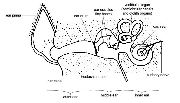
Image credit: Sunshineconnelly at en.wikibooks, CC BY 3.0 via Wikimedia Commons
The sympathetic nerve, cranial nerve VII (facial nerve), and cranial nerve VIII (vestibulocochlear nerve) all pass through the middle and inner ear structures. Infections that extend through these areas can affect these nerves and cause:
- Anisocoria (unequal pupil size via sympathetic nerve involvement)
- Facial paresis / paralysis
- Vestibular signs (head tilt, ataxia or wobbly gait)
- Decreased facial sensation (if cranial nerve V is involved)
- Tetraparesis (with intracranial extension / brain abscess formation)
Diagnosis: Imaging and Myringotomy
MRI is the gold standard for evaluating otitis media / interna when brainstem involvement is suspected. MRI clearly visualizes soft tissue structures in the brain (including abscesses), which are often obscured on CT scans due to bone artifact. However, CT scans can still play a role in making the diagnosis — they’re often more accessible, faster, and provide excellent detail of bony structures like the tympanic bullae.
Another essential diagnostic tool is myringotomy, a procedure in which a needle is passed through the ear canal and eardrum to sample middle ear fluid. This allows for cytology and culture testing to identify infectious organisms. It's important to realize, however, that not all cases yield positive cultures. In cats, for example, up to 50% of cases are sterile and related to allergies rather than infections.
⚠️ Note: Myringotomy ruptures the eardrum, so topical ear medications should not be used until the eardrum has healed (usually 2 to 4 weeks) - even in the face of an infection.
Treatment recommendations depend on the severity and progression of the disease.
Medical Therapy
- Steroids to control inflammation (even in patients diagnosed with an infection) - e.g. prednisone or prednisolone
- Antibiotics, ideally based on culture, for a minimum of 2 months
Patients should be closely monitored for signs of recurrence or worsening (which could indicate spreading of infection into the brain).
Medical therapy alone may not fully resolve an infection, particularly when a patient has intracranial extension. Therefore, surgery may be recommended to establish drainage.
Surgical Intervention
- Ventral bulla osteotomy (VBO) is the most common surgical approach
- Involves accessing the middle ear through the underside of the skull and surgically creating a hole
- Allows for drainage and removal of infectious material
- Helps prevent further intracranial spread
Postoperative complications can include nerve deficits that lead to a transient Horner’s Syndrome or facial paralysis (even if not present preoperatively). These complications are usually temporary and owners must be prepared for changes in their pet's appearance or facial function.
Allergy: A Root Cause Often Overlooked
Interestingly, a significant portion (up to 50%) of otitis media / interna cases in cats are caused by allergies rather than infection. Allergies can cause chronic inflammation and set the stage for middle ear disease. It’s also not unusual for cats to develop otitis media / interna in one ear and subsequently develop the same problem in the contralateral ear within a year or two if the underlying allergy is not addressed. Bilateral cases are also common in cats with allergies.
Dermatologic evaluation and allergy management are essential components of long-term care. Without it, the cycle of ear infections and potential neurologic complications is likely to continue.
Key Takeaways
- Neurologic symptoms can range from subtle to serious in pets with middle or inner ear infections.
- Neurological signs like anisocoria, ataxia, and facial paralysis may signal a deeper issue like extension of infection into the brain (brain abscess).
- MRI and myringotomy are powerful tools for diagnosis; culture results may be negative in up to 50% of feline cases.
- Treatment often requires long-term antibiotics, and in advanced cases, surgery is crucial to stop the infection from spreading to the brain.
- If allergies are the cause then allergy management is vital for preventing recurrence and bilateral involvement.